Conditions

What I treat — and what to know about it.

A plain-language guide to the colorectal and general surgical conditions seen most often at the practice. These notes are for orientation; nothing here replaces a personal consultation.

Colorectal cancer

The most common cancer in Malaysian men. Found early, it is one of the most curable.

Colorectal cancer — lesions in the bowel shown in detail against the full digestive system
Two of the most common sites at which colorectal cancer develops — the rectum and the sigmoid colon.

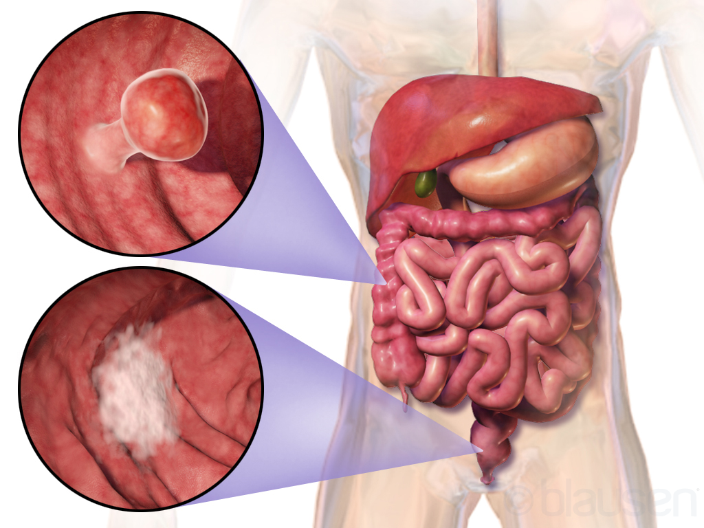
Colorectal cancer develops, in almost all cases, from a small benign growth in the bowel called a polyp. Polyps grow silently and slowly over many years. If they are found early — at colonoscopy — they can be removed in the same sitting and the cancer is prevented before it ever starts. When polyps are not found, a small minority eventually become invasive cancer.

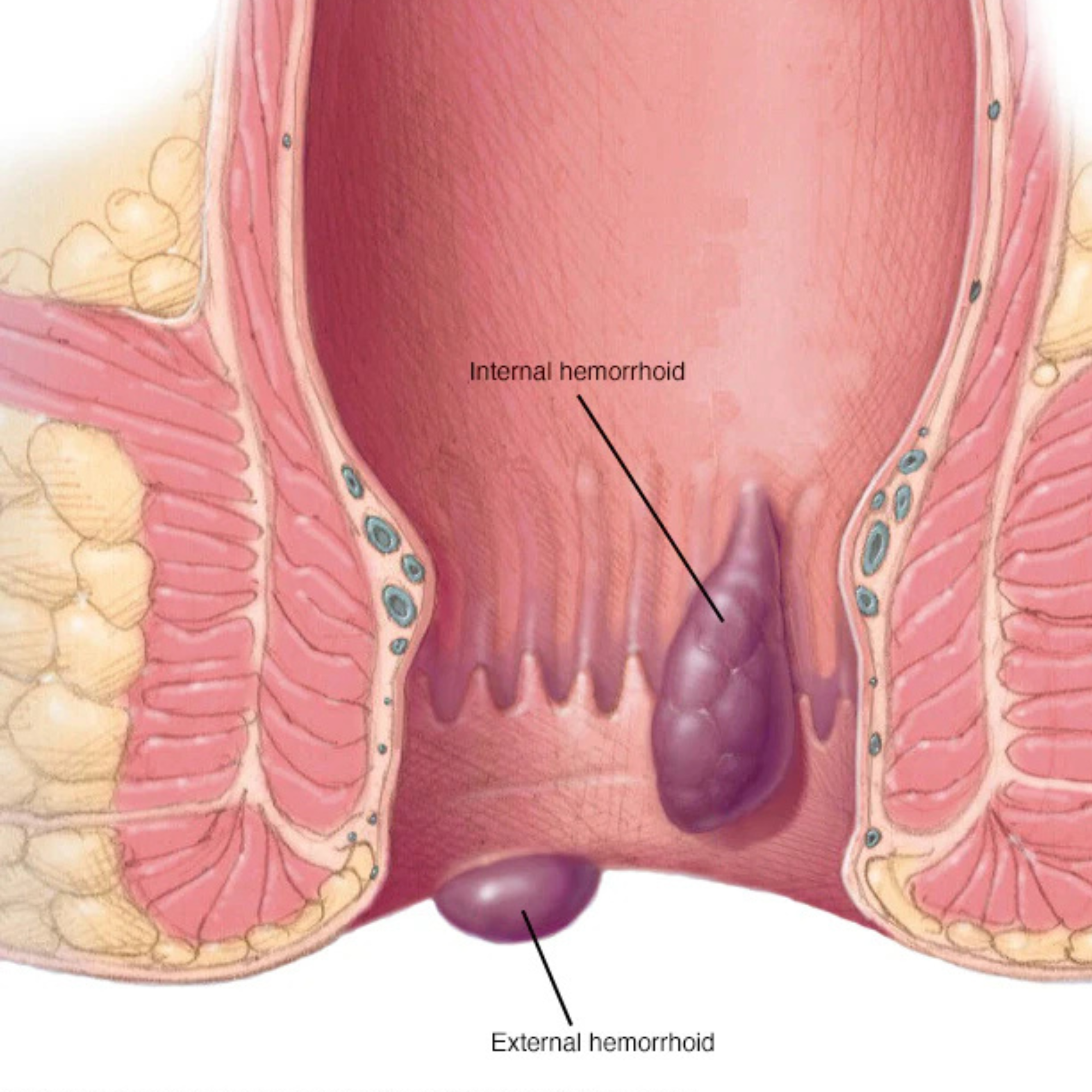
Early colorectal cancer rarely produces dramatic symptoms. Quietly persistent changes are more typical: a change in the shape or frequency of bowel movements, a sense of incomplete emptying, intermittent rectal bleeding (often blamed, wrongly, on haemorrhoids), unexplained iron-deficiency anaemia, or a low-grade abdominal discomfort. Weight loss and severe symptoms tend to appear later — which is why screening matters.

Treatment depends heavily on the stage of disease at diagnosis. Early-stage cancers are often curable with surgery alone, increasingly through minimally invasive keyhole approaches. More advanced cancers are managed with a combined plan — chemotherapy, radiotherapy and surgery — coordinated across a multidisciplinary team. By a long way, the most important variable in outcome is the stage at which it is found.

When to see a specialist

Any new rectal bleeding, a persistent change in bowel habit beyond a few weeks, unexplained tiredness or anaemia, or a family history of bowel cancer. None of these symptoms guarantee cancer — most have benign explanations — but they justify a single consultation to settle the question.

Haemorrhoids Buasir

Common, manageable, and almost never the social embarrassment patients fear.

Internal and external haemorrhoids — swollen blood vessels in the lower rectum and anus
Internal and external haemorrhoids — swollen blood vessels of the anorectum.

Haemorrhoids are swollen blood vessels in the lower rectum and anus. More than half of adults will experience them at some point. They are not dangerous — but they are frequently uncomfortable, and a quiet source of anxiety for patients across Kajang and the surrounding suburbs, many of whom delay seeking care for years.

Typical symptoms include bright red bleeding noticed on toilet paper or in the bowl during a bowel movement, itching, a sense of incomplete emptying, and in more advanced cases a lump that protrudes during defecation. Some haemorrhoids cause sudden severe pain when they thrombose — the swollen vessel clots and becomes tender.

About eighty percent of cases do not require surgery. Adequate dietary fibre, water, topical creams and improved toilet habits resolve most symptoms. For grade 2 and 3 haemorrhoids that do not settle, in-clinic options include rubber band ligation (a quick, almost painless office procedure) and laser haemorrhoidoplasty. Surgical excision is reserved for the most severe grade 4 disease.

A note on rectal bleeding

Bleeding is the most common reason patients first attend a colorectal clinic. It is almost always benign — a haemorrhoid or a fissure — but identical bleeding can be the only sign of early bowel cancer. A short, dignified examination usually settles the question in a single visit.

Anal fissure

A small tear, a disproportionately sharp pain — usually treatable without an operation.

An anal fissure — a small tear in the lining of the anal canal
The small tear in the anal lining that produces such disproportionately sharp pain.

An anal fissure is a small split in the lining of the anal canal, most often caused by the passage of a hard or large stool. The characteristic symptom is a sharp, tearing pain at the moment of bowel motion, often followed by an aching discomfort that can last hours. A small amount of bright red blood on the toilet paper is common.

Acute fissures usually heal within weeks with stool-softening measures, increased fluid and fibre, and topical medications that relax the anal sphincter. Chronic fissures — those persisting beyond six to eight weeks — sometimes require a small day-case procedure, such as a botulinum toxin injection or a lateral internal sphincterotomy, to break the cycle of pain and spasm.

Most patients return to normal within a short course of treatment. The condition can recur with constipation; the single most useful long-term measure is bowel-habit conditioning — avoiding constipation, avoiding straining, and not lingering on the toilet.

Anal fistula

An abnormal tract that almost always requires surgical correction.

Anal fistula — abnormal tunnel between the anal canal and the perineal skin
The fistula tract — running from the anal canal to the skin of the perineum.

An anal fistula is a small abnormal tunnel that develops between the inside of the anal canal and the skin of the perineum. It almost always originates as an infection in one of the small glands that drain into the anal canal — the gland becomes blocked, forms an abscess, and the abscess eventually drains through the skin, leaving the tunnel behind.

Patients usually present with recurrent boil-like swellings, intermittent discharge of pus or blood, or persistent moisture and irritation. The diagnosis is clinical, supported in complex cases by an MRI of the pelvis to map the fistula tract.

Fistulas do not heal spontaneously and are not safely managed by repeated drainage alone. Surgical correction is required. Several techniques exist — fistulotomy, seton placement, advancement flaps, LIFT, and laser ablation — each chosen according to the tract's anatomy and the patient's continence. The aim is to clear the disease while protecting the function of the anal sphincter.

Rectal prolapse

When the rectum slides outside the body — distressing, but surgically correctable.

Bowel anatomy showing the lower rectum — the segment that can prolapse through the anal canal
The lower bowel and rectum — the segment that can protrude through the anus in rectal prolapse.

Rectal prolapse describes the protrusion of the rectum through the anus. It can range from a small internal slide noticed only on imaging, to a full-thickness external prolapse that becomes visible after every bowel movement. It is more common in older women, in patients with chronic straining, and in those with pelvic-floor weakness following childbirth.

Symptoms include the sensation or sight of a protrusion, mucus discharge, bleeding, and varying degrees of faecal incontinence as the prolapse stretches the sphincter complex over time. It is a quality-of-life condition: rarely dangerous, often deeply distressing.

Surgical correction is offered laparoscopically in most cases — typically a ventral mesh rectopexy or a resection rectopexy — to restore the rectum to its normal anatomical position. The approach is selected to match the patient's age, fitness, and the specific anatomy of the prolapse.

Inflammatory bowel disease

Crohn's disease and ulcerative colitis. A lifelong condition that benefits from a steady surgical partnership.

Ulcerative colitis and Crohn's disease — the two main types of inflammatory bowel disease, with their characteristic inflammation patterns
Ulcerative colitis (left) involves continuous inflammation of the colon. Crohn's disease (right) can affect any part of the digestive tract in patches.

Inflammatory bowel disease (IBD) is the umbrella term for two chronic inflammatory conditions of the gut: Crohn's disease, which can affect any part of the digestive tract; and ulcerative colitis, which affects only the colon and rectum. Both are lifelong diseases, marked by periods of remission interrupted by flares of inflammation.

Medical therapy — anti-inflammatories, immunosuppressants and biologics — is the foundation of management and is led by a gastroenterologist. Surgery is reserved for specific situations: complications such as strictures, fistulas or perforations in Crohn's disease; severe medically-refractory ulcerative colitis; and the long-term cancer risk that comes with extensive colitis.

For ulcerative colitis, the definitive operation is panproctocolectomy with an internal pouch (IPAA) — the entire colon and rectum are removed, and a J-shaped pouch is fashioned from the small bowel to restore bowel continuity without a permanent stoma. It is technically demanding surgery, performed at the practice laparoscopically.

Irritable bowel syndrome IBS

A common functional disorder — uncomfortable, fluctuating, but not dangerous. Managed medically, not surgically.

Irritable bowel syndrome (IBS) is a functional disorder of the gut in which the bowel is structurally normal but behaves abnormally — typically alternating between constipation and diarrhoea, with abdominal cramping, bloating, and a sense of incomplete evacuation. It affects perhaps one in ten adults and is one of the most common reasons patients are referred for colorectal review.

The role of a specialist consultation is twofold: first, to confirm the diagnosis by excluding structural disease — particularly in patients above fifty, or where there are alarm features such as rectal bleeding, weight loss, or a family history of bowel cancer. A colonoscopy is often the test that settles the question. Second, to set a sensible management plan: dietary measures (often a structured low-FODMAP trial), stress management, targeted medication for the dominant symptom, and reassurance.

IBS is a lifelong condition that comes and goes. The aim is to control symptoms and rule out anything more serious — surgery has no role.

Diverticular disease

Small pouches in the colon wall that become inflamed or infected — common, often manageable, occasionally surgical.

Diverticula are small out-pouchings of the colon wall, most often in the sigmoid colon. They are extremely common with age — perhaps half of adults above sixty have them, often without ever knowing. The trouble starts when one becomes inflamed (diverticulitis) or infected, producing left lower abdominal pain, fever, change in bowel habit and sometimes bleeding.

Most flares of uncomplicated diverticulitis settle with antibiotics and dietary measures. Recurrent attacks, complications (abscess, perforation, fistula formation) or persistent bleeding may require surgical resection of the affected segment — usually laparoscopically.

When to see a specialist

Severe abdominal pain with fever, persistent bleeding, or two or more flares of diverticulitis. A colonoscopy is usually done once the acute episode has settled, to map the extent of disease and exclude other pathology.

Constipation

Difficulty passing stool — usually managed by simple measures, but persistent cases deserve a specialist look.

Constipation is one of the most common digestive complaints, and one of the most often dismissed. It can mean infrequent bowel movements, hard stool, a sense of incomplete evacuation, or excessive straining. The everyday causes are well known — inadequate fibre and water, sedentary lifestyle, certain medications, and the modern habit of ignoring the urge.

Most patients respond fully to a structured plan: 25–35 g of dietary fibre daily, adequate fluids, regular exercise, and a consistent toilet routine. Where these measures fail, or when constipation is new in an adult over fifty, accompanied by bleeding, weight loss or change in stool calibre — a specialist consultation and colonoscopy is warranted to exclude obstructive disease.

Obstructed defaecation syndrome

Difficulty emptying the rectum despite the urge — a pelvic-floor problem, not a simple constipation.

Obstructed defaecation syndrome (ODS) is a specific form of constipation in which the patient feels the urge to pass stool but cannot effectively empty the rectum. Symptoms include prolonged, repeated straining; a sensation of incomplete evacuation; the need to support the perineum or vagina with a finger to evacuate; and frequent small, fragmented bowel movements.

The underlying cause is usually mechanical — internal rectal prolapse (intussusception), rectocele (a bulge of the rectum into the vagina), enterocele, or pelvic-floor dyssynergia (the muscles failing to relax in coordination). Diagnosis often requires specialised investigations such as defaecating proctography or pelvic-floor MRI.

Treatment is staged: pelvic-floor physiotherapy and biofeedback first, dietary measures, and where structural problems persist, surgery — typically a laparoscopic ventral mesh rectopexy or STARR procedure. It is a quality-of-life condition that is often under-treated because patients assume nothing can be done.

Solitary rectal ulcer syndrome SRUS

A rare but well-recognised cause of rectal bleeding and mucus discharge — almost always linked to straining.

Solitary rectal ulcer syndrome (SRUS) is an uncommon but distinctive condition in which one or more ulcers form on the inner lining of the rectum, usually on the anterior wall a few centimetres above the anus. Despite the name, the ulcer is not always solitary. Typical symptoms are rectal bleeding, mucus discharge, a sense of incomplete evacuation, and a feeling of obstruction during bowel movements.

The condition is closely linked to chronic straining and to internal rectal prolapse, which causes the rectal wall to trap itself during defaecation. Diagnosis is by colonoscopy with biopsy — important because the appearance can resemble cancer or inflammatory bowel disease.

First-line treatment is behavioural: stop straining, treat constipation, biofeedback retraining of the pelvic floor. Where internal prolapse is the underlying driver, surgical correction (rectopexy) may be needed. The condition responds well to a structured approach but recovery is slow — months rather than weeks.

Perianal abscess

A collection of pus near the anus — almost always a surgical emergency requiring prompt drainage.

A perianal abscess is an acute collection of pus in the tissues around the anus, usually arising from infection of one of the small anal glands. The presentation is unmistakable: a hot, painful, tender swelling near the anus, often with fever and difficulty sitting. Pain is severe and worsens over hours.

Treatment is urgent surgical drainage — antibiotics alone are not enough. The procedure is short, performed under anaesthetic, and provides immediate relief. A significant proportion of perianal abscesses develop into a chronic anal fistula over the following months; surveillance and definitive fistula surgery may be needed later.

Don't wait

An untreated perianal abscess can progress to severe sepsis. If you suspect one — a painful, swelling lump near the anus that is rapidly worsening — present to a hospital or specialist clinic the same day.

Gallstones

Common, often silent. A good example of when surgery is straightforward and rewarding.

Gallstones anatomy — gallbladder, bile duct, pancreas and small intestine
The gallbladder sits beneath the liver and drains via the bile duct into the small intestine.

Gallstones are hardened deposits — most often cholesterol — that form within the gallbladder, a small pouch beneath the liver that stores bile. Many people carry gallstones for years without symptoms; these silent stones usually require no treatment. The picture changes when a stone obstructs the outflow of the gallbladder, causing biliary colic — a characteristic right-upper abdominal pain that often radiates to the back or shoulder, frequently after fatty meals.

Symptomatic gallstones do not improve with diet or medication; they require surgical removal of the gallbladder (cholecystectomy). The operation is performed laparoscopically through four small abdominal cuts, usually as a day-case or single overnight stay. Patients return to office work within a week and to normal activity within two to three weeks.

A small number of patients develop complications — gallstone pancreatitis, cholangitis, or stones lodged in the bile duct — which require more urgent and slightly more complex management. These are usually identified on imaging before surgery and dealt with in coordination with a gastroenterologist.

Acute pancreatitis

Sudden inflammation of the pancreas — almost always caused by gallstones or alcohol. Most cases settle, some are life-threatening.

Acute pancreatitis is the sudden inflammation of the pancreas, the gland that produces digestive enzymes and insulin. The two most common causes by a wide margin are gallstones (a small stone passes down the bile duct and irritates the pancreatic duct) and heavy alcohol intake. Symptoms are unmistakable: severe upper-abdominal pain radiating to the back, persistent vomiting, and a fever — pain often so severe that patients lean forward to relieve it.

Most cases are mild and settle with hospital admission, intravenous fluids, pain relief, and time. A small but significant minority become severe — with multi-organ involvement and a real mortality risk. Diagnosis is by blood tests (elevated amylase or lipase) and imaging.

Once the acute episode resolves, the underlying cause must be addressed. For gallstone pancreatitis, this means laparoscopic cholecystectomy — usually during the same admission or within a few weeks. Without removal of the gallbladder, the risk of a second, often worse, attack is high.

Don't dismiss the pain

Severe upper-abdominal pain that wraps around to the back, persistent vomiting, or any abdominal pain after a heavy meal in someone with known gallstones — present to a hospital the same day. Pancreatitis is one of the diagnoses for which delay genuinely changes the outcome.

Gastritis

Inflammation of the stomach lining — extremely common, very treatable, and rarely needs surgery.

Gastritis — inflammation and ulceration of the stomach lining
Inflammation of the stomach lining, sometimes with ulceration, producing upper-abdominal discomfort and nausea.

Gastritis is inflammation of the stomach lining. It is one of the most common upper-gastrointestinal complaints seen in Malaysian clinics, with most cases linked to Helicobacter pylori infection, regular use of anti-inflammatory medications (NSAIDs), alcohol, or chronic stress. The typical symptoms are upper-abdominal discomfort, nausea, a sense of fullness after small meals, and occasionally ulceration of the stomach lining.

Most gastritis is diagnosed clinically and confirmed where needed by an OGD (upper endoscopy) with a biopsy taken to test for H. pylori. Treatment is medical: acid-suppressing medication, eradication therapy for H. pylori where present, dietary advice, and addressing modifiable contributors. Surgery has no routine role in uncomplicated gastritis.

When to seek review

Persistent upper-abdominal pain, vomiting, dark-coloured stools, unexplained weight loss, or anaemia — all reasons for a same-week specialist review. An OGD typically settles the question in a single appointment.

GERD Gastro-oesophageal Reflux Disease

Chronic acid reflux — heartburn and regurgitation that, over time, can damage the oesophagus.

Closed and open sphincter — the mechanism of gastro-oesophageal reflux disease
A competent sphincter (left) versus the open, refluxing sphincter (right) seen in GERD.

Gastro-oesophageal reflux disease (GERD) describes the chronic backflow of stomach contents into the oesophagus. The lower oesophageal sphincter — the muscular valve at the base of the oesophagus — becomes weakened or relaxes inappropriately, allowing acid to wash up. The result is heartburn, regurgitation, a sour taste in the mouth, and over years the risk of changes to the oesophageal lining.

Most patients are managed medically — acid-suppressing medications, weight management, dietary advice, and avoiding late evening meals. A diagnostic OGD is performed when symptoms are persistent, when there are alarm features such as weight loss or difficulty swallowing, or in patients above fifty as part of cautious workup.

Surgery is reserved for a small minority — those with severe, medically-resistant reflux, or with complications such as a large hiatal hernia. Anti-reflux surgery is performed laparoscopically.

Hernia

A defect in the abdominal wall — best repaired electively, before complications develop.

Hernia repair — abdominal wall defect with surgical port positions
An incisional hernia of the abdominal wall, with the keyhole port positions used for laparoscopic mesh repair.

A hernia is a weakness or gap in the abdominal wall through which the abdominal contents push out, producing a visible bulge. They are extremely common in adults of all ages, and most produce some discomfort — particularly on standing, lifting or coughing. Hernias do not heal by themselves and tend to enlarge slowly over time.

The reason for repair is rarely cosmetic; it is to remove a small but real lifetime risk of an emergency — incarceration or strangulation of bowel within the hernia, which is a surgical emergency. Repair is performed laparoscopically in most cases, with a small soft mesh placed to reinforce the abdominal wall from inside. Most patients return to office work within a week.

The main types

Inguinal hernia — by far the commonest, presenting as a bulge in the groin. More common in men. May extend into the scrotum if neglected.

Femoral hernia — also in the groin, sitting slightly below the inguinal canal. Less common, but with a higher risk of strangulation, particularly in older women. Generally repaired sooner rather than later.

Umbilical / para-umbilical hernia — at or just beside the navel. Very common, often noticed after pregnancy, weight gain, or as a long-standing childhood hernia that has enlarged.

Incisional hernia — develops through the scar of a previous abdominal operation, sometimes years later. Repair is more complex because the abdominal wall is already weakened; a larger mesh is usually needed.

Appendicitis

The classical surgical emergency — diagnosed clinically, treated promptly.

Appendicitis — inflamed appendix shown in body context with magnified view
The inflamed appendix at the start of the large bowel, with a magnified view of the swollen tissue.

Acute appendicitis is inflammation of the appendix, a small finger-like projection at the start of the large bowel. The presentation is classical: pain that begins around the navel and shifts to the right lower abdomen over several hours, with nausea, loss of appetite and a mild fever. Diagnosis is largely clinical, often supported by ultrasound or CT scan.

Once diagnosed, the appendix is removed promptly — almost always laparoscopically, through three small cuts. Most patients are discharged home within twenty-four to forty-eight hours and resume normal activity within a fortnight.

Untreated appendicitis carries a real risk of perforation and intra-abdominal sepsis, which is why timing matters. Any patient with progressive right-lower abdominal pain should be assessed urgently in a hospital setting.

Lumps, bumps & cysts

Skin and subcutaneous lumps — the bulk of everyday general surgery. Most are benign and removed in a single appointment.

The single largest category of general surgery work is the assessment and removal of skin and subcutaneous lumps. This includes lipomas (soft fatty lumps), sebaceous cysts (smooth, slow-growing skin cysts), fibromas, enlarged lymph nodes, and a wide variety of less common lesions. Most are entirely benign and produce no problem beyond cosmetic concern, friction with clothing, or occasional inflammation.

The clinical role of a specialist is twofold. First, to confirm clinically — and where indicated, with ultrasound or biopsy — that the lump is benign. Second, where removal is requested or warranted, to excise it cleanly with minimal scarring. Most procedures are done as a day-case under local anaesthetic, taking thirty minutes or less. Larger lesions, deep cysts, or anything in a cosmetically sensitive area may need short general anaesthetic.

Abscesses are a separate category — collections of pus that develop suddenly, are hot and painful, and require prompt incision and drainage rather than elective excision. Where a lump is suddenly painful, growing rapidly, or developing skin changes, same-week review is sensible.

When to have a lump assessed

Any lump that is new, growing, painful, firm, fixed to underlying tissue, or associated with skin changes deserves a single specialist visit. Most are benign and quickly settled. The minority that aren't are caught early — which is the whole point.

Frequently asked

Common questions about these conditions.

What are the early signs of haemorrhoids (buasir)?

The most common early sign is painless bright-red bleeding noticed on toilet paper, in the bowl, or coating the stool. Itching, a feeling of incomplete emptying, mucus discharge, and a soft lump that may protrude during a bowel movement and reduce on its own are also typical. Severe pain is unusual unless a haemorrhoid has thrombosed (clotted).

When should I see a colorectal surgeon for rectal bleeding?

See a specialist if rectal bleeding lasts more than two weeks, is associated with a change in bowel habit (looser stools, constipation, or thinner stools) lasting more than three weeks, is accompanied by abdominal pain, weight loss, fatigue or anaemia, or if you are 45 years or older and have never had a colonoscopy. Rectal bleeding should never be assumed to be just haemorrhoids without evaluation.

What is the difference between an anal fissure and a haemorrhoid?

An anal fissure is a small tear in the lining of the anal canal that causes sharp, often severe pain during and after a bowel movement, sometimes with a small amount of bright-red bleeding. A haemorrhoid is a swollen vascular cushion that usually bleeds painlessly and may protrude. Pain is the key distinguisher — fissures hurt sharply, internal haemorrhoids typically do not.

What is inflammatory bowel disease (IBD)?

IBD is a group of chronic conditions that cause inflammation of the digestive tract, most commonly Crohn's disease and ulcerative colitis. Symptoms include persistent diarrhoea, rectal bleeding, abdominal pain, weight loss and fatigue. IBD is distinct from IBS (irritable bowel syndrome). Diagnosis is made by colonoscopy with biopsies and imaging.

Does a hernia always need surgery?

Not always. Small, asymptomatic hernias can sometimes be monitored. However, hernias do not resolve on their own and most progress over time. Surgery is recommended if the hernia is painful, enlarging, restricting activity, or if there is any risk of incarceration or strangulation (a surgical emergency).

Have a concern from this list?

A single consultation is usually enough to settle the question — confirm the diagnosis, exclude what matters, and plan the next step.

Request an Appointment