Procedures

Minimally invasive surgery, described properly.

The colorectal and general procedures performed by Dr. Qisti at KPJ Kajang Specialist Hospital. Each entry includes the indication, the technique, and what to expect from preparation through to recovery.

A note on technique

Why minimally invasive surgery is the default.

Almost every operation listed on this page can be performed either through a traditional open incision or by keyhole. The choice of approach is not a matter of fashion. Smaller incisions mean less pain, less blood loss, shorter hospital stays, faster return to work, and lower wound-complication rates. For pelvic surgery, the precision of laparoscopic technique matters most.

The constraint, always, is the safety of the operation. There are situations — extensive adhesions, locally advanced cancer, sudden bleeding — in which open surgery remains the correct choice. The aim is to do the right operation, in the right way, for the right patient.

Dr. Nik Qisti Fathi performing laparoscopic surgery in the operating theatre at KPJ Kajang Specialist Hospital
Colorectal

Colorectal Surgery

Eight procedures across keyhole, transanal and anorectal technique — including bowel resection, stoma formation, and minor anorectal procedures.

01

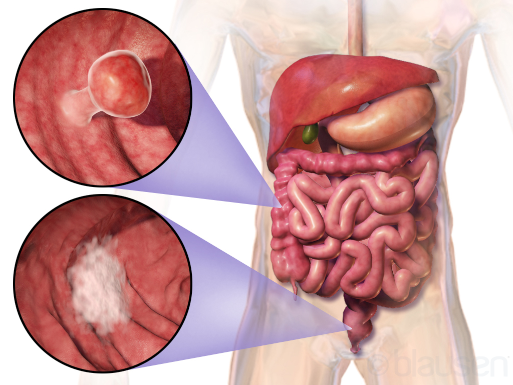
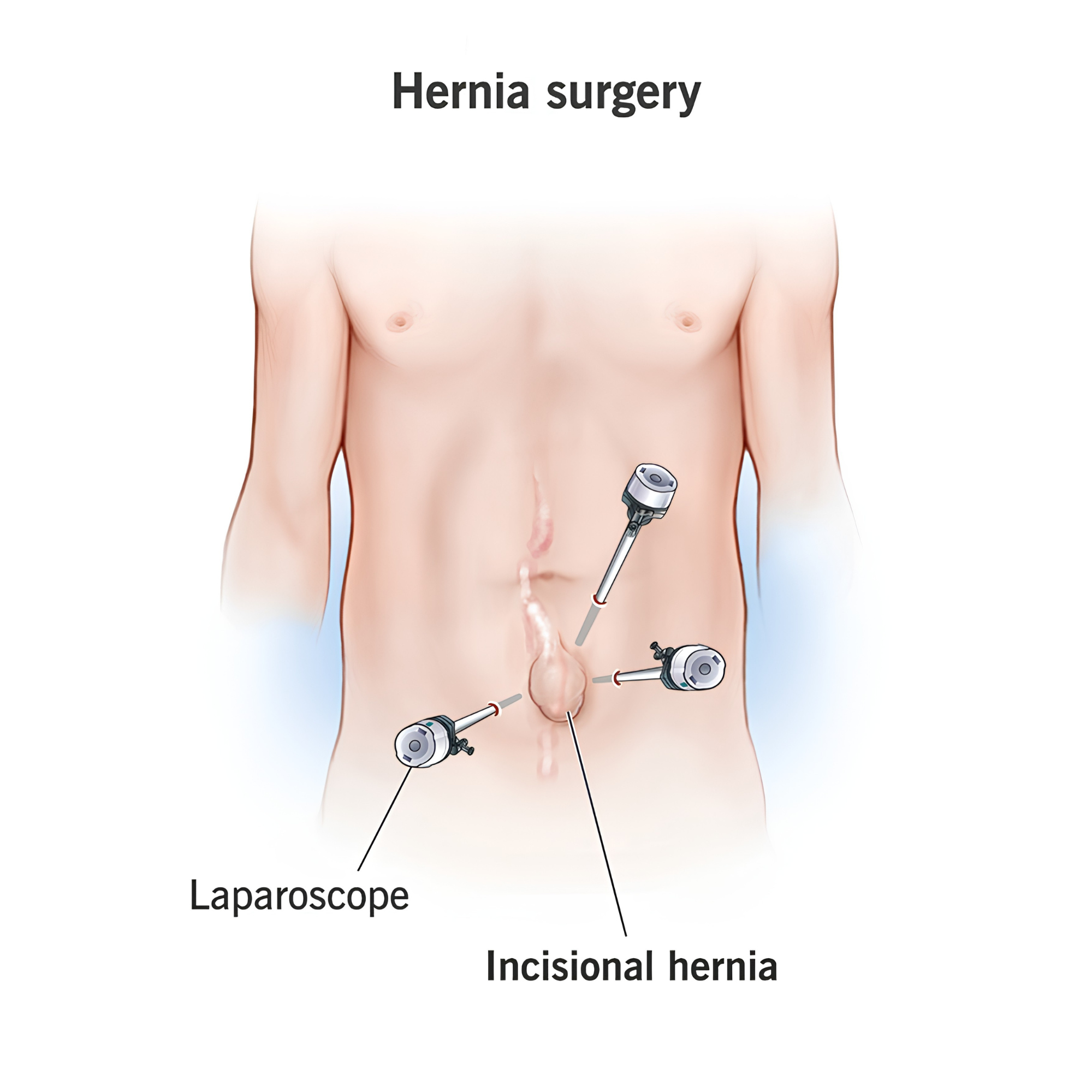
Laparoscopic Colorectal Cancer Surgery

Keyhole bowel resection with full lymph-node clearance.

The keyhole removal of a segment of the bowel containing a cancer, together with its draining lymph nodes. Performed through four or five small abdominal incisions, with one slightly larger cut to deliver the specimen.

The technique reduces post-operative pain, shortens hospital stay to typically four to six days, and allows most patients to return to office work within three to four weeks. The same oncological principles apply as in open surgery — the resection is no less radical because it is performed laparoscopically.

Approach
Laparoscopic
Hospital stay
4 – 6 days
Back to work
3 – 4 weeks
Colorectal cancer — common sites of disease in the bowel
02

Laparoscopic Anterior Resection

Removal of the upper rectum with bowel reconnection.

The keyhole removal of the upper part of the rectum — performed for rectal cancer, or for benign disease such as diverticulitis. After the diseased segment is taken out, the bowel is rejoined inside the pelvis, preserving the natural route of bowel evacuation.

For lower rectal cancers, a temporary protective stoma is sometimes fashioned to allow the new join to heal — this is typically reversed three to four months later. The aim throughout is bowel preservation: avoiding a permanent stoma wherever it is safely possible.

Approach
Laparoscopic
Hospital stay
5 – 7 days
Stoma
Often temporary
The bowel before and after anterior resection — diseased segment removed, bowel ends rejoined
03

Panproctocolectomy + IPAA (J-Pouch)

Complete bowel removal with internal pouch reconstruction — no permanent stoma.

One of the most technically demanding operations in colorectal surgery. The entire colon and rectum are removed — usually for severe ulcerative colitis or familial polyposis — and the last loop of small bowel is folded into a J-shaped reservoir (ileal pouch–anal anastomosis) and joined to the anus.

The result is a permanent solution to disease affecting the entire large bowel, without a permanent external stoma. The pouch takes several months to mature, and a temporary protective ileostomy is usually used during this period.

Approach
Laparoscopic
Hospital stay
7 – 10 days
Pouch matures
3 – 6 months
Panproctocolectomy with ileal pouch-anal anastomosis — the J-pouch reconstruction
04

Transanal Minimally Invasive Surgery TAMIS

Rectal tumours removed through the anus — no abdominal incisions.

A specialised technique for the removal of large rectal polyps and selected early rectal cancers — performed entirely through the anus, with no abdominal incisions. A small operating platform is placed in the anal canal, and the lesion is excised under direct vision with carbon dioxide insufflation.

TAMIS is well-suited to lesions that would otherwise require a major bowel resection but are still confined to the rectal wall. Patient selection is critical; not every rectal lesion is a candidate.

Approach
Transanal
Hospital stay
1 – 2 days
Back to work
1 – 2 weeks
Endoscopic removal of a rectal lesion using a snare — the principle behind TAMIS
05

Laparoscopic Right Hemicolectomy

Keyhole removal of the right colon — among the quickest recoveries.

The keyhole removal of the right side of the large bowel — performed most often for cancer of the caecum or ascending colon, and occasionally for benign disease such as a large right-sided polyp. The bowel ends are rejoined inside the abdomen.

Recovery is among the quickest of the major colorectal operations. Most patients are eating normally by the third day, home within four to five days, and back to office work within three weeks.

Approach
Laparoscopic
Hospital stay
4 – 5 days
Stoma
Not required
Right hemicolectomy — the right colon and the planned line of resection
06

Haemorrhoid Treatment

Banding, laser or excision — matched to the grade of disease.

Treatment is matched to the grade of the disease. Grade 1 and 2 haemorrhoids are usually managed in clinic with rubber band ligation — a brief, painless office procedure that takes minutes. Grade 3 disease responds well to laser haemorrhoidoplasty, a minimally invasive day-case procedure that uses a precise laser fibre to coagulate and shrink the haemorrhoidal tissue from within.

Surgical haemorrhoidectomy — the traditional excision — is reserved for severe grade 4 disease. It is the most definitive treatment, but also the one with the longest recovery; the post-operative weeks are uncomfortable, and adequate analgesia is planned in advance.

Options
Banding · Laser · Excision
Hospital stay
Same-day
Recovery
Days to weeks
Rubber band ligation of an internal haemorrhoid — the ligator device places a band at the base of the haemorrhoid
07

Lateral Internal Sphincterotomy & Botox Injection

Day-case treatments for chronic anal fissure that doesn't settle with medication.

Chronic anal fissures that resist conservative treatment are often locked in a cycle of pain and sphincter spasm — the muscle tightens because of pain, the tightness prevents healing, and the cycle continues. Two short procedures break this cycle.

Botox injection — a small dose of botulinum toxin is injected into the internal anal sphincter, temporarily relaxing it for two to three months. This is long enough for the fissure to heal in most patients. No incision, day-case, sometimes performed in clinic. Reversible.

Lateral internal sphincterotomy — a precise, controlled division of a small portion of the internal sphincter to relieve the spasm permanently. Performed under anaesthetic as a day-case. High success rate; a small risk of altered continence is discussed in advance.

Approach
Day-case anorectal surgery
Hospital stay
Same day
Recovery
1 – 2 weeks
Anal fissure — anatomy and treatment with botox injection and lateral internal sphincterotomy
08

Laparoscopic Stoma Creation

Keyhole formation of a temporary or permanent stoma — for IBD, obstruction, or as part of complex pelvic surgery.

A stoma is a surgically created opening that allows bowel contents to be diverted to the abdominal wall, where they are collected in a discreet appliance. Stomas are sometimes temporary (to protect a healing bowel join after surgery) and sometimes permanent (where the bowel below cannot be used).

Formed laparoscopically through small incisions in the abdomen, the procedure is part of a larger surgical plan — most commonly during colorectal cancer surgery, severe inflammatory bowel disease, complex anal fistula management, or to relieve large-bowel obstruction. Patients are supported by a specialist stoma care nurse before and after surgery; modern stoma appliances are unobtrusive and allow a normal life.

Approach
Laparoscopic
Hospital stay
3 – 5 days
Reversibility
Temporary stomas reversed in 3 – 6 months
Laparoscopic stoma creation
Dr. Qisti reviewing patient notes at his clinic
General

General Surgery

Four procedures covering the most common general surgical conditions seen in the practice — cholecystectomy, hernia, appendix and the broad category of skin lumps.

09

Laparoscopic Cholecystectomy

Keyhole gallbladder removal — usually a day-case.

The keyhole removal of the gallbladder — the operation for symptomatic gallstones. Performed through four small cuts, typically as a day-case or single overnight stay. There are no long-term dietary restrictions afterwards; the liver continues to produce bile, which drains directly into the intestine.

Approach
Laparoscopic
Hospital stay
Day-case / 1 night
Back to work
7 – 10 days
Laparoscopic gallbladder removal — surgical instruments and laparoscope positions
10

Hernia Repair — Laparoscopic IPOM & TEP

Keyhole repair of all four common hernia types, using the technique matched to the defect.

Hernia repair is performed laparoscopically in most cases through three small abdominal incisions, with a soft mesh placed to reinforce the abdominal wall. The specific technique is matched to the type of hernia:

Laparoscopic IPOM repair (intraperitoneal onlay mesh) — the technique used for umbilical, paraumbilical, and incisional hernias. The mesh is placed inside the abdomen against the inner wall, covering the defect from within.

Laparoscopic TEP repair (totally extraperitoneal) — the technique used for inguinal, femoral, and obturator hernias of the groin. The mesh is placed in a plane outside the peritoneum, avoiding the abdominal cavity entirely. Particularly well-suited to bilateral inguinal hernias and to recurrent hernias.

Approach
Laparoscopic (TEP / TAPP)
Hospital stay
Day-case
Back to work
1 – 2 weeks
Laparoscopic incisional hernia repair — laparoscope and instrument ports
11

Appendicectomy

Laparoscopic removal of an inflamed appendix.

Laparoscopic removal of an inflamed appendix — the standard treatment for acute appendicitis. Performed through three small cuts, typically with discharge within twenty-four to forty-eight hours. Where the appendicitis has progressed to perforation or abscess, a slightly longer admission may be needed.

Approach
Laparoscopic
Hospital stay
1 – 2 days
Back to work
1 – 2 weeks
Appendicectomy — inflamed appendix shown in body context with magnified view
12

Lumps, Bumps & Cyst Excision

Day-case removal of lipomas, sebaceous cysts and benign skin lumps. Drainage of abscesses.

The bulk of everyday general surgical work: removing benign skin and subcutaneous lumps — lipomas, sebaceous cysts, fibromas, enlarged lymph nodes — and draining abscesses that present acutely. Most procedures are performed as day-case under local anaesthetic and take thirty minutes or less. Larger or deeper lesions, and anything in a cosmetically sensitive area, may need a brief general anaesthetic.

Histology of the removed tissue is sent routinely. Where a lesion is unusual or suspicious, the result determines whether any further treatment is required.

Approach
Local or short general anaesthetic
Hospital stay
Day-case
Back to work
Same day to 1 week
Lumps, bumps and cyst excision
Diagnostic endoscopy at KPJ Kajang Specialist Hospital
Diagnostic

Diagnostic Procedures

Four diagnostic procedures — colonoscopy, upper-GI endoscopy, dedicated anorectal imaging, and keyhole abdominal investigation — that support and extend the surgical practice.

13

Colonoscopy lower-GI endoscopy

The most important diagnostic and preventive procedure in colorectal medicine. Dedicated full guide on a separate page.

A colonoscopy is a slim flexible endoscope passed through the entire large bowel, used both to investigate symptoms (bleeding, change in bowel habit, anaemia, abdominal pain) and to screen for bowel cancer in patients above 45. It is the single most useful test in modern colorectal medicine — the only one that simultaneously diagnoses and treats by removing polyps in the same sitting.

Performed under conscious sedation by an anaesthetist. The procedure itself takes 20–30 minutes; total appointment time is a half-day from admission through to discharge.

Approach
Endoscopic, under sedation
Duration
20 – 30 minutes
Recovery
Same-day discharge

Read the full colonoscopy guide

Colonoscopy anatomy — flexible endoscope traversing the large bowel
14

Upper-GI Endoscopy OGD / Gastroscopy

Diagnostic endoscopy of the upper digestive tract — the camera test for the oesophagus, stomach and duodenum.

An upper-GI endoscopy — known clinically as an OGD (oesophagogastroduodenoscopy) or simply a gastroscopy — is a short outpatient procedure in which a slim flexible camera is passed through the mouth to examine the upper digestive tract. It is the standard investigation for persistent heartburn, upper abdominal pain, vomiting, swallowing difficulty, anaemia, and suspected stomach ulcers. Biopsies — including for H. pylori — can be taken in the same sitting.

Performed under conscious sedation by an anaesthetist; the procedure itself takes 10–15 minutes. Often combined with a colonoscopy on the same day where both ends of the digestive tract need to be assessed.

Approach
Endoscopic, under sedation
Duration
10 – 15 minutes
Recovery
Same-day discharge
Upper-GI endoscopy (OGD / gastroscopy) — the scope is passed through the mouth to examine the oesophagus, stomach and duodenum
15

Endo-anal Ultrasound

Detailed ultrasound imaging of the anal sphincters and surrounding tissues — for fistula mapping and sphincter assessment.

A specialised ultrasound probe is gently inserted into the anal canal to produce high-resolution images of the anal sphincter muscles, surrounding fat, and any abnormal tracts (such as fistulas). It is the investigation of choice for mapping complex anal fistulas before surgery, assessing sphincter integrity after childbirth or previous surgery, and characterising perianal masses.

Brief outpatient procedure — typically 10–15 minutes, no sedation needed, no preparation beyond a normal bowel movement on the day. Findings are discussed immediately.

Approach
Outpatient ultrasound
Duration
10 – 15 minutes
Sedation
Not required
Endo-anal ultrasound
16

Diagnostic Laparoscopy

A keyhole look inside the abdomen — when imaging hasn't given a clear answer and a definitive diagnosis is needed.

Diagnostic laparoscopy is a short keyhole procedure in which a camera is introduced into the abdomen through one or two small incisions, allowing direct visualisation of the abdominal and pelvic organs. It is used where imaging (CT, ultrasound, MRI) has been inconclusive and a confident diagnosis is needed to plan further treatment.

Common indications include unexplained chronic abdominal pain, suspected appendicitis in women where the diagnosis is unclear, staging of upper-GI or pelvic malignancy, assessment of peritoneal disease, and investigation of unexplained ascites. Where a problem is identified during the procedure, definitive treatment can sometimes be carried out in the same sitting.

Approach
Laparoscopic, under general anaesthetic
Hospital stay
Day-case or 1 night
Recovery
3 – 7 days
Diagnostic laparoscopy

Considering one of these procedures?

A specialist consultation will clarify whether surgery is needed, which approach suits your case, and what to expect from preparation through to recovery.

Request an Appointment